
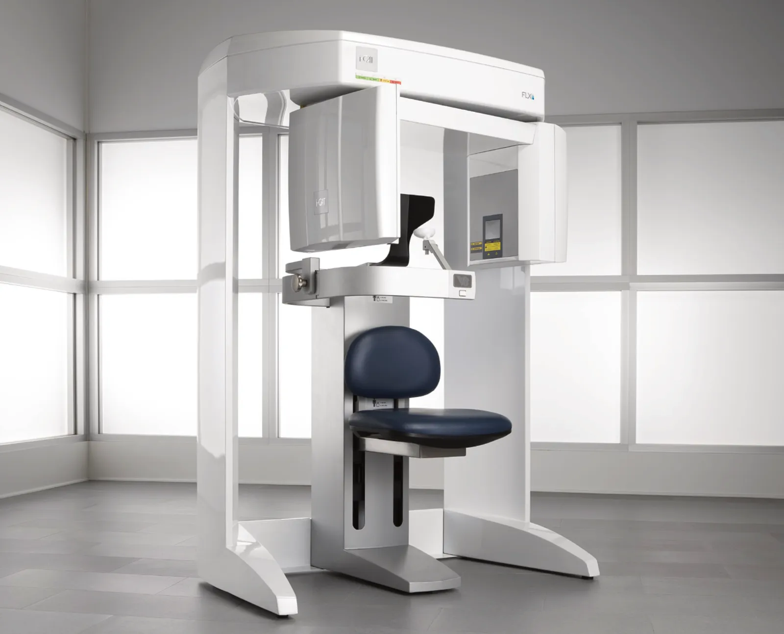
QuickScan+ cuts dose and scan time to just 4.8 seconds, producing a full 3D dentition at a radiation dose comparable to a panoramic image, with reconstruction in under 30 seconds
Made by us
Dental Equipment › Dental Imaging
WEB-030
i-CAT FLX V-Series
i-CATThe i-CAT FLX V-Series cone beam 3D imaging product family offers a flexible, upgradable 3D solution designed to help practices grow.
- Sensor type
- Amorphous Silicon Flat Panel Sensor with CsI Scintillator
- Grayscale resolution
- 16-bit
- Voxel size
- .4 mm, .3 mm, .25 mm, .2 mm, .125 mm
- Collimation
- Electronically controlled, fully adjustable
Overview
What it does.
The i-CAT FLX V-Series cone beam 3D imaging product family offers a flexible, upgradable 3D solution designed to help practices grow. Providing optimal clarity, ease-of-use and control, the system's field-of-view can be upgraded based on the types of procedures the practice performs, enabling more precise diagnoses and better surgical predictability. As the first upgradable dedicated CBCT unit, the V-Series combines highly precise cone beam 3D technology with procedure-based upgradable fields-of-view, software, and flexible planning and treatment tools. It is especially beneficial for implants, restorations, oral and maxillofacial surgery, orthodontics, periodontics, endodontics, and TMJ and airway analysis.
Key features
What makes it different.
PureScan advanced image technology for i-CAT's clearest 3D and 2D images
SmartScan STUDIO touchscreen interface with guided, customizable workflow (preset scan options or Quickpicks for field-of-view, dose, and resolution)
i-PAN feature for traditional 2D panoramic images using the same high-quality 3D sensor
Ergonomic Stability System (ESS) for seated patient positioning, minimizing movement and unnecessary retakes
Widely adjustable 3D fields-of-view through electronically controlled collimation (V8, V10, or V17 models)
Tx STUDIO treatment planning software (Mac + Windows compatible), developed with Anatomage: implant planning, surgical guides, 3D cephalometric analysis, airway analysis, virtual studies, impressionless models, face-MATCH (photo superimposition), AnatoModel (bone/teeth segmentation)
Open software architecture, integrating with orthodontic programs, CAD/CAM systems, imaging software, and practice management programs
STL file export compatible with a wide variety of CAD/CAM and 3D-printing systems
Automatic nerve canal tracing to check for root entanglement prior to extractions
Volumetric airway analysis: automatic computation of total airway volume with color-coded constriction values
Less than 90 seconds from scan to plan; complete workup capturable in under 10 seconds
Surgical guides available through Anatomage service, delivered in under a week
Voted Best Cone Beam CT Scanner (Dental Townie Choice Awards); Pride Institute Best of Class Technology Award (2013–2015); Top 100 Dental Products Report
i-CAT University: ongoing education via webinars, regional classes, on-site training, Users' Meetings, and 3D Congress
Installed in more than 6,500 sites worldwide, with telephone support, remote assistance, and field service teams
Technical specifications
The numbers.
| Specification | Value |
|---|---|
| Sensor type | Amorphous Silicon Flat Panel Sensor with CsI Scintillator |
| Grayscale resolution | 16-bit |
| Voxel size | .4 mm, .3 mm, .25 mm, .2 mm, .125 mm |
| Collimation | Electronically controlled, fully adjustable |
| Scan time | 4.8, 8.9, 14.7, 17.8, or 26.9 seconds |
| Exposure type | Pulsed |
| Field-of-view (V8) | 8x5 cm, 8x8 cm |
| Field-of-view (V10) | 8x5, 8x8, 16x4, 16x6, 16x8, 16x10 cm |
| Field-of-view (V17) | 8x5, 8x8, 16x4, 16x6, 16x8, 16x10, 16x11, 16x13, 23x17 cm |
| Reconstruction shape | Cylinder |
| Typical reconstruction time | Less than 30 seconds |
| Viewing and treatment software | Included |
| DICOM compatible | Yes |
| Unit size | 48" (W) x 69.5" (H) x 36.37" (D) |
| Patient position | Seated |
| Wheelchair accessible | Yes |
Documentation
Downloads.
Product enquiry
Plan your next step.
Explore this product with our team.