
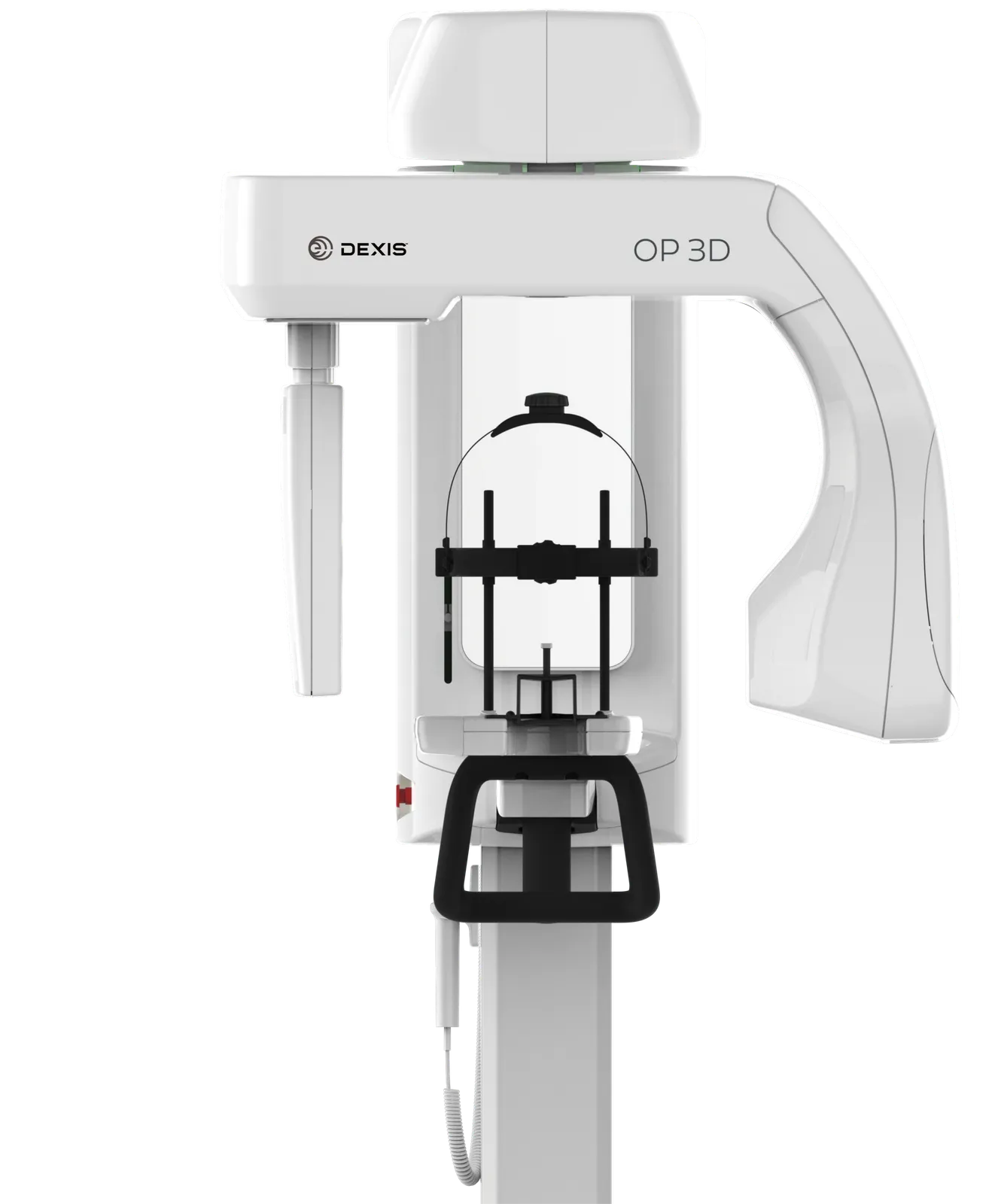
Fully upgradeable: choose to add cephalometric imaging, or fully upgrade to add 3D imaging
Made by us
Dental Equipment › Dental Imaging
WEB-028
Dexis ORTHOPANTOMOGRAPH OP 3D
DexisThe DEXIS OP 3D is a unit designed for advanced dental imaging needs.
- Image volume sizes (H x Ø)
- 5x5, 6x9, 9x11, 9x14 cm (optional); volume height/location adjustable via SMARTVIEW 2.0
- Image receptor
- CMOS
- Pixel size (sensor & image)
- 99 μm
- Tube voltage
- 60 - 90 kV
Overview
What it does.
The DEXIS OP 3D is a unit designed for advanced dental imaging needs. It is a complete X-ray platform that provides easy-to-use features throughout the entire dental imaging workflow. The versatile panoramic, cephalometric and 3D programs offer image excellence for a variety of users ranging from general dental practitioners to maxillofacial surgeons. It respects the legacy of the ORTHOPANTOMOGRAPH product family while renewing the meaning of image quality, efficiency and ease-of-use. The OP 3D imaging unit replaces lead typically used for tubehead radiation shielding designs with a more ecological and environmentally friendly alternative that provides equivalent radiation attenuation, and its power save feature reduces overall energy consumption of the practice.
Key features
What makes it different.
9-second standard and pediatric panoramic scan times, providing clear image definition with fewer movement artifacts and lower patient dose
ORTHOfocus feature: automatically obtains the optimum panoramic image layer, enabling forgiving patient positioning and consistent image quality
Multilayer pan: optionally supplies five layers within one exposure at the same scan time and dosage as a single panoramic exposure
Bitewing-like view as a quick alternative to intraoral bitewing imaging
ORTHOselect user interface: intuitive selection of imaging area (individual teeth, upper/lower jaw, or TMJ), with automatic optimum FOV
SMARTVIEW 2.0 user-interface: two-dimensional scout images before CBCT examination, with adjustable FOV location and height, supporting ALARA radiation protocols
QUICKcompose feature for fast preview of panoramic, cephalometric, and 3D images
Metal Artifact Reduction (MAR), active across all FOV sizes, aiding endodontics, implants, and maxillofacial imaging
Edge Preserving Noise Reduction (EPNR) filter, smoothing noise while preserving sharp edges and clinical detail
Variety of resolutions: Low Dose Technology (LDT), Standard, High resolution, and Endo resolution (80 μm voxel, available for 5x5 FOV)
ORTHOceph Plus design (cephalometric): patented design keeps correct height for CEPH imaging after a panoramic scan; dedicated X-ray source for high capacity and minimal patient dose
Cephalometric protocols: lateral and pediatric lateral projections (adjustable field width), posterior-anterior (PA) projections, carpus imaging (optional holder)
Easy wheelchair accessibility
Supports implantology, endodontics, impactions, TMJ, trauma, and periodontics
Technical specifications
The numbers.
| Specification | Value |
|---|---|
| Image receptor | CMOS |
| Pixel size (sensor & image) | 99 μm |
| Tube voltage | 60 - 90 kV |
| Tube current | 2 - 16 mA |
| Scan time | 9 s |
| Image field height | 147 mm |
| Imaging programs | Standard, Segmented, Pediatric, Lateral TMJ, Bitewing |
| Image receptor | CMOS |
| Pixel size (sensor & image) | 99 μm |
| Tube voltage | 60 - 95 kV |
| Tube current | 2 - 14 mA |
| Scan time | 10.5 and 8.1 s |
| Image field height | 180 - 223 mm |
| Image field width | 160 - 260 mm |
| Imaging programs | Lateral and Pediatric Lateral (adjustable field width), Posterior-Anterior (PA), Carpus* |
| Image detector | CMOS |
| Image voxel size | 80 - 400 μm |
| Tube voltage | 95 kV |
| Tube current | 2 - 12.5 mA |
| Scan time | 10 - 20 s |
| Image volume sizes (H x Ø) | 5x5, 6x9, 9x11, 9x14 cm (optional); volume height/location adjustable via SMARTVIEW 2.0 |
| Tube focal spot | 0.5 IEC 336 (IEC 60336/2005) |
| DICOM support | Available as a software option |
Documentation
Downloads.
Product enquiry
Plan your next step.
Explore this product with our team.