
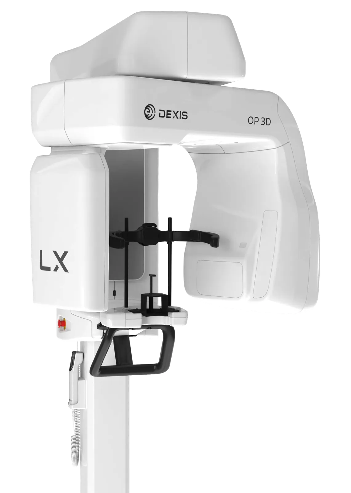
Flexible FOV, from 5x5 up to 15x20 complete high-resolution scans, with 96 additional customizable options and four resolutions (including endo mode and low dose mode)
Made by us
Dental Equipment › Dental Imaging
WEB-027
Dexis ORTHOPANTOMOGRAPH OP 3D LX
DexisBuilt on OP 3D technology, the OP 3D LX is the next generation of proven DEXIS cone beam technology — a multimodality imaging platform that expands 3D diagnostic capabilities with a wide range of clinical applications supporting evolving practices and enhanced diagnostic confidence.
- HU capacity
- 35 kJ, 49,000 HU
- Weight
- 120 kg / 265 lbs
- Weight
- 155 kg / 342 lbs
- Image volume sizes (HxD)
- 50x50, 60x90, 80x80, 100x100, 120x150, 150x200 mm
Overview
What it does.
Built on OP 3D technology, the OP 3D LX is the next generation of proven DEXIS cone beam technology — a multimodality imaging platform that expands 3D diagnostic capabilities with a wide range of clinical applications supporting evolving practices and enhanced diagnostic confidence. The 2D and 3D imaging options cover a wide range of dental extraoral needs, from endodontics to complex surgical cases. This next-generation system offers flexible FOV options ranging from 5x5 up to 15x20 — the largest view option available on a DEXIS OP 3D platform to date. With short scan times, the OP 3D LX captures the maxillofacial complex and large diagnostic areas in one non-stitched scan for fast workflows.
Key features
What makes it different.
Scalable solution: standard panoramic, extraoral bitewing and 3D imaging up to 10x10, upgradable to 12x15 and 15x20 volume sizes or cephalometric modality
Consistent high-quality images via implant planning tools and noise reduction filters minimizing artifacts and noise
Intuitive user interface for patient positioning, 3D/panoramic/cephalometric area selection, and instant X-ray preview without opening viewing software
Stitch-free scans: single-pass capture with no stitching on every scan size
Re-engineered head support, optimized for orthodontic and surgical applications without interfering with patient's soft tissue profile
Next-generation automated ICE (Implant Contrast Enhancer) and MAR (Metal Artifact Reduction) for greater visibility of internal metal structures
New ergonomic exposure switch with light indicator (ready/exposing) and adjustable device/remote sounds
AI-assisted workflow features in DTX Studio Clinic: automatic focal trough, tooth-centric navigation, patient positioning correction, auto 3D tooth positioning, mandibular nerve canal annotation, merge of CBCT and intraoral scans
Automatic Dose Control (ADC): automatically measures patient anatomy and sets exposure values for optimal image quality and dose
Supports endodontics, maxillofacial surgery, orthodontics, TMJ analysis, airway analysis, periodontics, prosthodontics, and implant placement (single unit, All-on-4®, zygomatic)
Optional 10-year warranty coverage
Wheelchair accessible
Technical specifications
The numbers.
| Specification | Value |
|---|---|
| Focal spot | 0.5 (IEC 60336/2020) |
| Tube voltage | 60 - 95 kV |
| Tube current | 2 - 16 mA |
| HU capacity | 35 kJ, 49,000 HU |
| Minimum total filtration | 3.4 mm Al @ 95 kV |
| Wheelchair accessible | Yes |
| Min. room height | 2100 - 2455 mm |
| Image detector | IGZO TFT |
| Sensor/image pixel size | 95 μm |
| Scan/exposure time | 1.4 - 15.0 s (Standard Pan 9.0 s) |
| Image field height | 128.4 - 187.0 mm |
| Imaging programs | Standard, HD Pan, segmented standard, pediatric, segmented pediatric, bitewing, lateral TMJ |
| Weight | 120 kg / 265 lbs |
| Image detector | IGZO TFT |
| Sensor/image pixel size | 95 μm |
| Scan/exposure time | 6.9 - 11.3 s |
| Image field height | 180.0 - 235.9 mm |
| Imaging programs | Lateral, pediatric lateral, PA, carpus |
| Weight | 155 kg / 342 lbs |
| Image detector | IGZO TFT |
| Image voxel size | 80 - 400 μm |
| Scan time | 8 - 30 s |
| Exposure time | 0.9 - 19.4 s |
| Image volume sizes (HxD) | 50x50, 60x90, 80x80, 100x100, 120x150, 150x200 mm |
| DICOM support | Yes |
| Width | 1345 mm / 53.0 in |
| Depth | 530 mm / 20.9 in |
| Length | 1100 mm / 43.3 in |
Documentation
Downloads.
Product enquiry
Plan your next step.
Explore this product with our team.